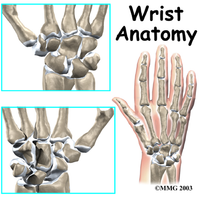
Wrist Anatomy
Introduction
Physical therapy in Asheville for Wrist
Welcome to Combined Therapy Specialties patient resource about Wrist problems.
The anatomy of the wrist joint is extremely complex, probably the most complex of all the joints in the body. The wrist is actually a collection of many bones and joints. These
bones and joints let us use our hands in lots of different ways. The wrist must be extremely mobile to give our hands a full range of motion. At the same time, the wrist must provide the strength for heavy gripping.
This guide will help you understand:
- what parts make up the wrist
- how those parts work together
#testimonialslist|kind:all|display:slider|orderby:type|filter_utags_names:Hand Pain|limit:15|heading:Hear from some of our patients who we treated for *Hand Pain*#
Important Structures
The important structures of the wrist can be divided into several categories. These include:
- bones and joints
- ligaments and tendons
- muscles
- nerves
- blood vessels
Bones and Joints
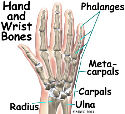
There are 15 bones that form connections from the end of the forearm to the hand. The wrist itself contains eight small bones, called:
Carpal Bones
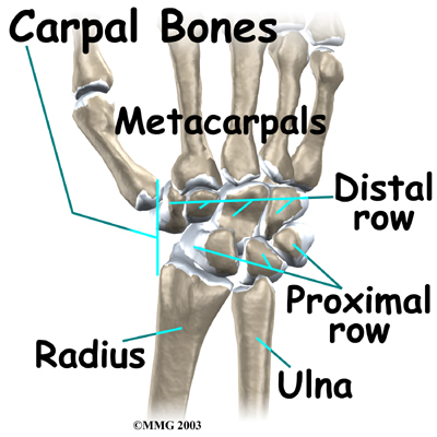
These bones are grouped in two rows across the wrist. The
Proximal Row
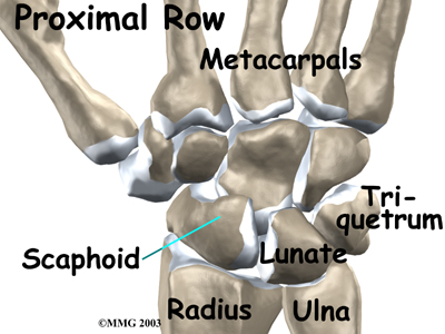
is where the wrist creases when you bend it. Beginning with the thumb-side of the wrist, the proximal row of carpal bones is made up of the scaphoid, lunate, and triquetrum. The second row of carpal bones, called the:
Distal Row
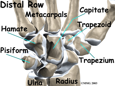
meets the proximal row a little further toward the fingers. The distal row is made up of the trapezium, trapezoid, capitate, hamate, and pisiform bones.
The proximal row of carpal bones connects the two bones of the forearm, the radius and the ulna, to the bones of the hand. The bones of the hand are called the
metacarpal bones. These are the long bones that lie within the palm of the hand. The metacarpals attach to the phalanges, which are the bones in the fingers and thumb.
One reason that the wrist is so complicated is because every small carpal bone forms a joint with the bone next to it. This means that what we call the wrist joint is actually made up of many small joints.
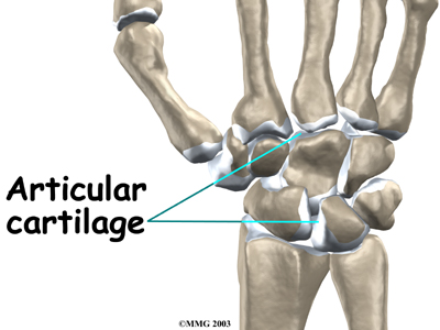 Articular cartilage is the material that covers the ends of the bones of any joint. Articular cartilage can be up to one-quarter of an inch thick in the large, weight-bearing joints. It is thinner in joints such as the wrist that don't support a lot of weight. Articular cartilage is white, shiny, and has a rubbery consistency. It is slippery, which allows the joint surfaces to slide against one another without causing any damage.
The function of articular cartilage is to absorb shock and provide an extremely smooth surface to make motion easier. We have articular cartilage essentially everywhere that two bony surfaces move against one another, or articulate. In the wrist, articular cartilage covers the sides of all the carpals and the ends of the bones that connect from the forearm to the fingers.
Ligaments and Tendons
Ligaments are soft tissue structures that connect bones to bones. The ligaments around a joint usually combine to form a:
Joint Capsule
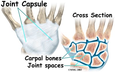
A joint capsule is a watertight sac that surrounds a joint and contains lubricating fluid called synovial fluid. In the wrist, the eight carpal bones are surrounded and supported by a joint capsule.
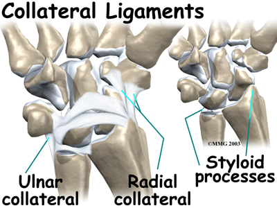 Two important ligaments support the sides of the wrist. These are the collateral ligaments There are two collateral ligaments that connect the forearm to the wrist, one on each side of the wrist.
As its name suggests, the ulnar collateral ligament (UCL) is on the ulnar side of the wrist. It crosses the ulnar edge (the side away from the thumb) of the wrist. It starts at the ulnar styloid, the small bump on the edge of the wrist (on the side away from the thumb) where the ulna meets the wrist joint. There are two parts to the cord-shaped UCL. One part connects to the pisiform (one of the small carpal bones) and to the transverse carpal ligament, a thick band of tissue that crosses in front of the wrist. The other goes to the triquetrum (a small carpal bone near the ulnar side of the wrist). The UCL adds support to a small disc of cartilage where the ulna meets the wrist. This structure is called the triangular fibrocartilage complex (TFCC) and is discussed in more detail below. The UCL stabilizes the TFCC and keeps the wrist from bending too far to the side (toward the thumb).
The radial collateral ligament (RCL) is on the thumb side of the wrist. It starts on the outer edge of the radius on a small bump called the radial styloid. It connects to the side of the scaphoid, the carpal bone below the thumb. The RCL prevents the wrist from bending too far to the side (away from the thumb).
Just as there are many bones that form the wrist, there are many ligaments that connect to and support these bones. Injury or problems that cause these ligaments to stretch or tear can eventually lead to arthritis in the wrist.
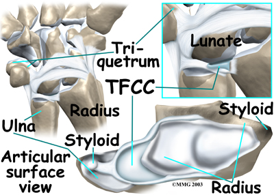 At the wrist, the end of the ulna bone of the forearm articulates with two carpal bones, the lunate and the triquetrum.
A unique structure mentioned earlier, the triangular fibrocartilage complex (TFCC), sits between the ulna and these two carpal bones.
The TFCC is a small cartilage pad that cushions this part of the wrist joint. It also improves the range of motion and gliding action within the wrist joint.
There are several important tendons that cross the wrist.
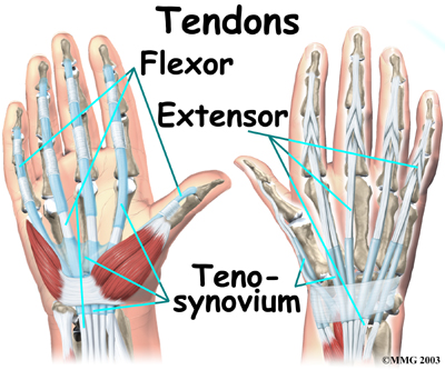 Tendons connect muscles to bone. The tendons that cross the wrist begin as muscles that start in the forearm. Those that cross the palm side of the wrist are the flexor tendons. They curl the fingers and thumb, and they bend the wrist. The flexor tendons run beneath the transverse carpal ligament (mentioned earlier). This structure lies on the palm side of the wrist. This band of tissue keeps the flexor tendons from bowing outward when you curl your fingers, thumb, or wrist. The tendons that travel over the back of the wrist, the extensor tendons, run through a series of tunnels, called compartments. These compartments are lined with a slick substance called tenosynovium, which prevents friction as the extensor tendons glide inside their compartment.
Muscles
The main muscles that are important at the wrist have been mentioned above in the discussion about tendons. These muscles generally start further up in the forearm. The tendons of these muscles cross the wrist. They control the actions of the fingers, thumb, and wrist.
Nerves
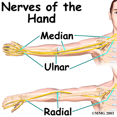
All of the nerves that travel to the hand cross the wrist. Three main nerves begin together at the shoulder: the radial nerve, the median nerve, and the ulnar nerve. These nerves carry signals from the brain to the muscles that move the arm, hand, fingers, and thumb. The nerves also carry signals back to the brain about sensations such as touch, pain, and temperature.
The
Radial Nerve
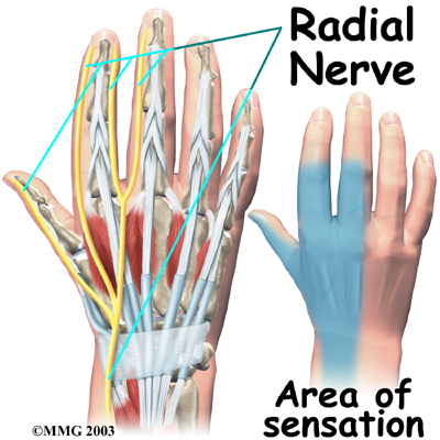
runs along the thumb-side edge of the forearm. It wraps around the end of the radius bone toward the back of the hand. It gives sensation to the back of the hand from the thumb to the third finger. It also goes to the back of the thumb and just beyond the main knuckle of the back surface of the ring and middle fingers.
Median Nerve
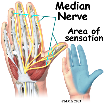
The Median Nurve travels through a tunnel within the wrist called the carpal tunnel. The median nerve gives sensation to the palm sides of the thumb, index finger, long finger, and half of the ring finger. It also sends a nerve branch to control the thenar muscles of the thumb. The thenar muscles help move the thumb and let you touch the pad of the thumb to the tips each of each finger on the same hand, a motion called opposition.
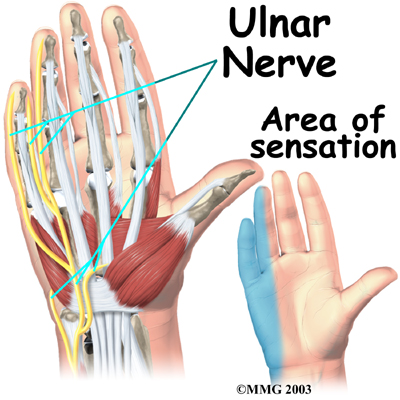 The ulnar nerve travels through a separate tunnel, called Guyon's canal. This tunnel is formed by two carpal bones (the pisiform and hamate), and the ligament that connects them.
After passing through the canal, the ulnar nerve branches out to supply feeling to the little finger and half the ring finger.
Branches of this nerve also supply the small muscles in the palm and the muscle that pulls the thumb toward the palm.
The nerves that travel through the wrist are subject to problems.
Constant bending and straightening of the wrist and fingers can lead to irritation or pressure on the nerves within their tunnels and cause problems such as pain, numbness, and weakness in the hand, fingers, and thumb.
Blood Vessels
Traveling along with the nerves are the:
Large Vessels
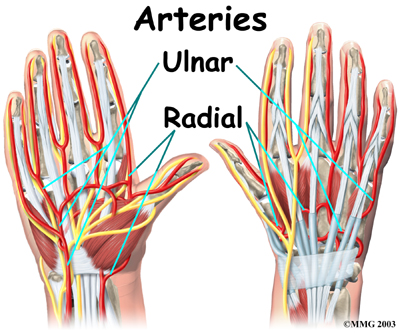
that supply the hand with blood. The largest artery is the radial artery that travels across the front of the wrist, closest to the thumb. The radial artery is where the pulse is taken in the wrist. The ulnar artery runs next to the ulnar nerve through Guyon's canal (mentioned earlier). The ulnar and radial arteries arch together within the palm of the hand, supplying the front of the hand and fingers. Other arteries travel across the back of the wrist to supply the back of the hand and fingers.
Summary
As you can see, the wrist is a complex area of the body. When you realize all the different ways we use our hands every day and all the different positions we put our hands in, it is easy to understand how hard daily life can be when the wrist doesn't work well.
Portions of this document copyright MMG, LLC.