Introduction
Physical therapy in Asheville for Shoulder
Welcome to Combined Therapy Specialties patient resource about Calcific Tendonitis of the Shoulder.
Calcific tendonitis of the shoulder happens when calcium deposits form on the tendons of your shoulder. The tissues around the deposit can become inflamed, causing a great deal of shoulder pain. This condition is fairly common. It most often affects people over the age of 40.
This article will help you understand:
- what happens in the shoulder with calcific tendonitis
- what tests your doctor will run to diagnose this condition
- what you can do to help relieve the pain.
#testimonialslist|kind:all|display:slider|orderby:type|filter_utags_names:Shoulder Pain|limit:15|heading:Hear from some of our patients who we treated for *Shoulder Pain*#
Anatomy
 Which part of the shoulder is affected?
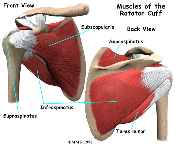
Calcific tendonitis occurs in the tendons (tendons attach muscles to bones) of the rotator cuff.
The is actually made up of several tendons that connect the muscles around your shoulder to the humerus (the larger bone of the upper arm).
Calcium deposits usually form on the tendon in the rotator cuff called the supraspinatus tendon.
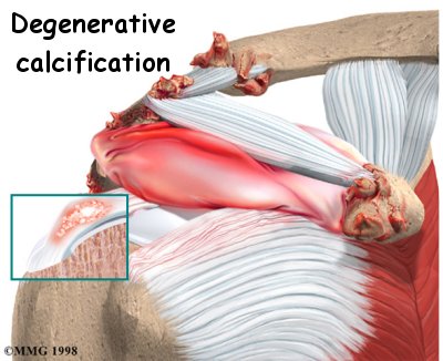
There are two different types of calcific tendonitis of the shoulder: degenerative calcification and reactive calcification. The wear and tear of aging is the primary cause of . As we age, blood flow to the tendons of the rotator cuff decreases. This makes the tendon weaker. Due to the wear and tear as we use our shoulder, the fibers of the tendons begin to fray and tear, just like a worn-out rope. Calcium deposits form in the damaged tendons as a part of the healing process.
Reactive calcification is different. Why it occurs is not clear. It doesn't seem to be related to degeneration, though it is more likely to cause shoulder pain than degenerative calcification. Doctors think of reactive calcification in three stages. In the pre-calcific stage, the tendon changes in ways that make calcium deposits more likely to form. In the calcific stage, calcium crystals are deposited in the tendons. Then they begin to disappear. The body simply reabsorbs the calcium deposits. Ironically, it is during this stage that pain is most likely to occur. In the post-calcific stage, the body heals the tendon, and the tendon is remodeled with new tissue.
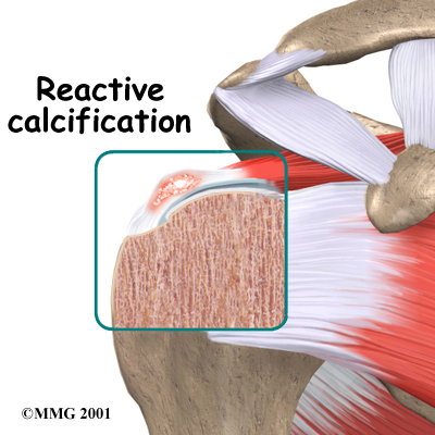
No one knows what triggers the body to reabsorb the deposits. But once this occurs and the tissue begins to be remodeled, the pain usually decreases or goes away altogether.
Related Document: A Patient's Guide to Shoulder Anatomy
Shoulder Anatomy Introduction
Causes
Why did I develop calcific tendonitis?
No one really knows what causes calcific tendonitis. Severe wear and tear, aging, or a combination of the two are involved in degenerative calcification. Some researchers think that calcium deposits form because there is not enough oxygen to the tendon tissues. Others feel that pressure on the tendons can damage them, causing the calcium deposits to form.
Reactive calcification is even more of a mystery. This type of problem occurs in younger patients and seems to go away by itself in many cases.
Symptoms
What tests will my doctor run?
While the calcium is being deposited, you may feel only mild to moderate pain, or even no pain at all. For some unknown reason, calcific tendonitis becomes very painful when the deposits are being reabsorbed. The pain and stiffness of calcific tendonitis can cause you to lose motion in your shoulder. Lifting your arm may become painful. At its most severe, the pain may interfere with your sleep.
Diagnosis
Your doctor will take a detailed medical history and do a thorough physical exam of your shoulder. The pain of calcific tendonitis can be confused with other conditions that cause shoulder pain. An X-ray is usually necessary to confirm the presence of calcium deposits. The X-ray will also help pinpoint the exact location of the deposits.
You will probably need to get several X-rays over time. This will help your doctor keep track of the changes in the amount of calcification. By following the changes in the calcium deposits, your doctor can determine whether the condition will heal by itself or perhaps require surgery.
Diagnosis
When you visit Combined Therapy Specialties, our physical therapist will take a detailed medical history and do a thorough physical exam of your shoulder. The pain of calcific tendonitis can often be confused with other conditions that cause shoulder pain.
Some patients may be referred to a doctor for further diagnosis. Once your diagnostic examination is complete, the physical therapists at Combined Therapy Specialties have treatment options that will help speed your recovery, so that you can more quickly return to your active lifestyle.
Our Treatment
Non-surgical Rehabilitation
When you begin your physical therapy program at Combined Therapy Specialties, our first goal will be to help control your pain and inflammation. Initial treatment is likely to be rest and anti-inflammatory medication, such as ibuprofen. The anti-inflammatory medicine is used mainly to control pain.
Our physical therapist may apply heat, ice or ultrasound treatments. Ultrasound has shown some benefit in reducing the size of the deposit and in helping people have less pain and better arm function. However, to get the full benefit, ultrasound treatments must be repeated often (up to 24 times) in a six-week period.
Your physical therapist can then create an individualized program of strengthening and stretching for your shoulder. It is very important to strengthen the muscles of the rotator cuff, as these muscles help control the stability of the shoulder joint. Strengthening these muscles can actually decrease the pressure on the calcium deposits in the tendon. Our physical therapist can also evaluate your workstation or the way you use your body when you do your activities and suggest changes. Simple changes in the way you sit or stand can ease pain and help you avoid further problems.
Post-surgical Rehabilitation
Rehabilitation after shoulder surgery can be a slow process. Although the time required for recovery varies, you will probably need to attend physical therapy sessions for about six to eight weeks, and should expect full recovery to take three to four months. Getting the shoulder moving as soon as possible is important. However, this must be balanced with the need to protect the healing tissues.
You may have to wear a sling to support and protect your shoulder for a few days after surgery. Our physical therapist may use ice and electrical stimulation treatments during your first few sessions to help control pain and swelling from the surgery. We may also use massage and other types of hands-on treatments to ease muscle spasm and pain.
Therapy can progress quickly after a simple arthroscopic resection. Our treatments start out with range-of-motion exercises and gradually work into active stretching and strengthening. You just need to be careful to avoid doing too much, too quickly.
Therapy goes slower after open surgery, where the shoulder muscles have been cut. Your physical therapist will usually wait up to two to three weeks before starting range-of-motion exercises. Exercises begin with passive movements. In passive exercises, our physical therapist will move your shoulder joint while your muscles stay relaxed. We will gently move your joint and gradually stretches your arm. Our physical therapist may also teach you how to do passive exercises at home.
Active therapy usually starts four to six weeks after surgery. Our physical therapist will instruct you on how to use your own muscle power in active range-of-motion exercises. We may have you begin with light isometric strengthening exercises. These exercises work the muscles without straining the healing tissues.
At about six weeks you start doing heavier strengthening. These exercises focus on improving the strength and control of the rotator cuff muscles and the muscles around your shoulder blade. Our physical therapist will help you retrain these muscles to keep the ball of the humerus in the socket. This helps your shoulder move smoothly during all your activities.
Some of the exercises we’ll teach you are designed get your shoulder working in ways that are similar to your work tasks and sport activities. Our physical therapist in Asheville will help you find ways to do your tasks that don't put too much stress on your shoulder. Before your physical therapy sessions end, our physical therapist will teach you a number of ways to avoid future problems.
Combined Therapy Specialties provides services for physical therapy in Asheville.
Physician Review
The pain of calcific tendonitis can often be confused with other conditions that cause shoulder pain. Your doctor will probably order an X-ray to confirm the presence of calcium deposits. The X-ray will also help pinpoint the exact location of the deposits.
You will probably need to get several X-rays over time. This will help your doctor keep track of the changes in the amount of calcification. By following the changes in the calcium deposits, your doctor can determine whether the condition will heal by itself or perhaps require surgery.
Your doctor may suggest a cortisone injection if your pain stays severe even after trying other nonsurgical treatments. Cortisone is a very powerful steroid. Cortisone can be very effective at temporarily easing inflammation and swelling.
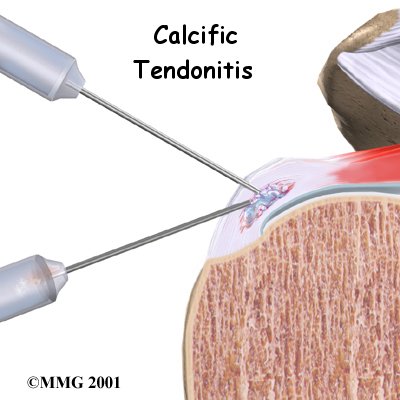
During the time when the calcium deposits are being reabsorbed, the pain can be especially bad. Your doctor may suggest trying to remove the calcium deposit by inserting two large needles into the area and rinsing with sterile saline. (Saline is simply a saltwater solution.) This procedure is called lavage. Sometimes lavage breaks the calcium particles loose. Then they can be removed with the needles. Getting rid of the calcium deposits can help speed up the healing. Even when lavage fails to remove calcium deposits, it may reduce pressure in the tendon, leading to less pain.
Shock wave therapy is a newer form of nonsurgical treatment. It uses a machine to generate shock wave pulses to the sore area. Patients generally receive the treatment once each week for up to three weeks. The impulses are thought to help break up the deposit so the body can more easily absorb it. Recent studies indicate that this form of treatment can help ease pain and reduce the size of the deposit.
Surgery
If the pain and loss of movement continue to get worse or interfere with your daily life, you may need surgery. Surgery for calcific tendonitis does not usually require patients to stay in the hospital overnight. It does require anesthesia.
Arthroscopic Resection
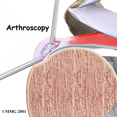
Most surgeries to correct calcific tendonitis of the shoulder are arthroscopic surgeries. The arthroscope is a special TV camera that can be inserted into the shoulder joint through a small incision in the skin. Other small incisions allow the surgeon to insert small surgical instruments into the joint as well. The surgeon uses the arthroscope to locate the calcium deposit in the rotator cuff tendon. Once the deposit is found, the surgeon uses the small instruments to resect (remove) the calcium deposits and rinse the area. Loose calcium crystals must be removed. They can be very irritating to the surrounding tissues.
Open Resection
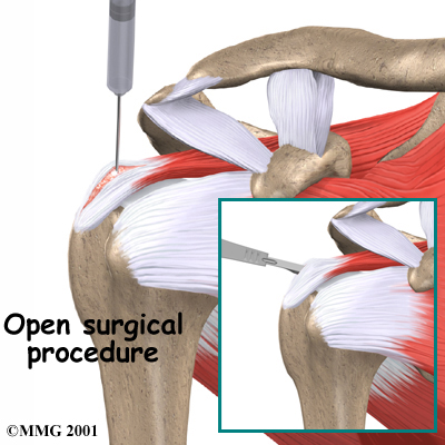
In rare instances, is necessary. In open surgery, the surgeon gets to the calcium deposit by cutting through muscles and other surrounding tissues. The tendon itself is cut to allow removal of the calcium deposits. The surgeon rinses the area to get rid of calcium crystals and then stitches the muscles and skin together.
Portions of this document copyright MMG, LLC.


 Which part of the shoulder is affected?


